A 3D reconstruction of AP view. The pelvic region of the trunk is the lower part of the trunk between the abdomen and the thighs.

Pelvis Anatomy Recon Orthobullets
The parietal pelvic fascia is removed to visualize the embedded autonomic pelvic nerves.

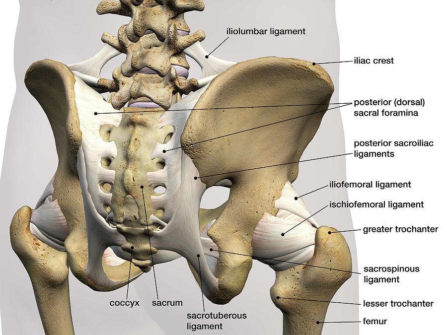
. De la prueba autor. The two pelvic bones are connected anteriorly by the pubic symphysis while posteriorly they articulate with the pelvic spine to form the sacroiliac joints. The anterior muscles posteriorly tilt the pelvis the posterior muscles anteriorly tilt the pelvis the muscles on the right side elevate the right side of the pelvis and therefore depress the left side of the pelvis and the muscles on the left side elevate the left side of the pelvis and therefore depress the right side of the pelvis.
From the quiz author. The pelvis plays several important functions in the human body. This is an online quiz called THS Anatomy Pelvis Posterior View.
Bony pelvis is formed posteriorly by the sacrum and the coccyx and laterally and. Medial view of a right-sided male hemipelvis. The sacrum and two innominate bones.
The bones of the pelvis and lower back work together to support the bodys weight anchor the abdominal and hip muscles and protect the delicate vital organs of the vertebral and abdominopelvic cavities. Anatomical landmarks within the vagina can be used to locate the position of such structures as the ureter and urethra and warn of their possible involvement in a vaginal laceration. The vertebral column of the lower back includes the five lumbar vertebrae the sacrum and the coccyx.
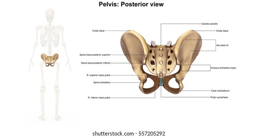
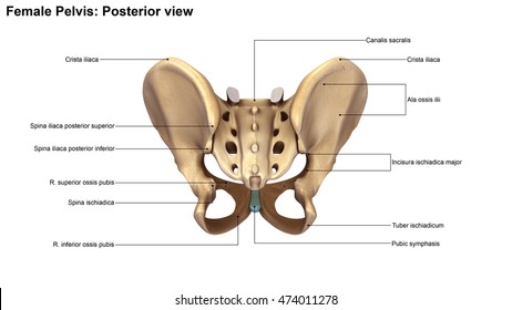
False and True Pelves. Roughened area located on the posterior medial side of the ilium of the hip bone. Bony pelvis or pelvic skeleton is formed by hip bones sacrum and coccyx.
The pelvic region is the area between the trunk or main body and the lower extremities or legs. Learn vocabulary terms and more with flashcards games and other study tools. The pelvic cavity and perineum.
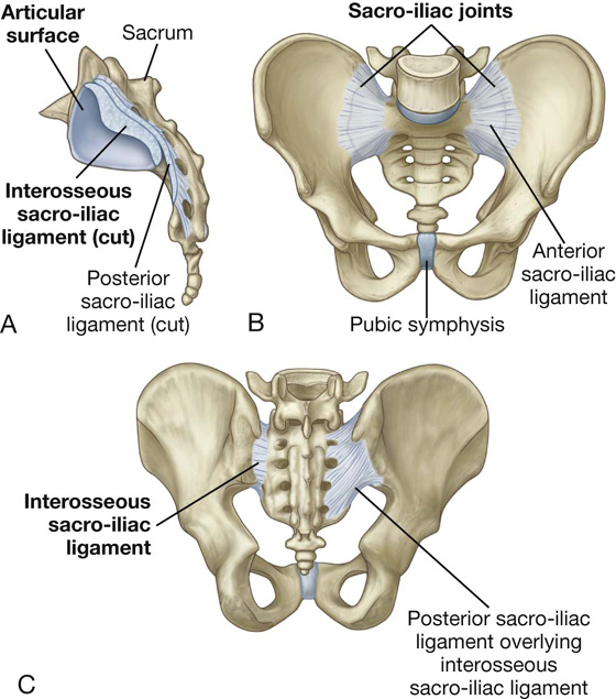
Bone And Ligaments Of Pelvis Posterior View. The three bones and three joints composing the pelvic ring have no inherent stability without vital ligamentous. Click on the tags below to find other quizzes on the same subject.
It has fibers that run almost laterally to connect the lateral posterior aspect of the upper two segments to the PSIS and the internal iliac crest. Start studying Pelvic Bone Anatomy. Major components of the bony pelvis frontal superior view of the female pelvis.
Features that most clearly distinguish the female from the male pelvis include a wider subpubic angle wider sciatic notch and greater distance from pubic symphysis and anterior. The true pelvis situated inferior to the caudal portion of the parietal peritoneum is considered the pelvic cavity Figure 41-3 and Box 41-1 The posterior wall of the pelvic cavity is formed by the sacrum and coccyx and the margins of the posterolateral wall are formed by the piriformis and coccygeus muscles Figure 41-4. There is a printable worksheet available for download here so you can take the quiz with pen and paper.
Identify the following parts of the pelvic girdle This quiz has tags. A cavity enclosed by the bones of the pelvis and containing the urinary bladder is the _________ cavity. Von make a Jell-O mold that has strawberries suspended in it and whipped cream on top the strawberries are ___________ while the whipped cream is.
Pelvic briminlet - a line from the sacral promontory to the upper part of the pubic symphysis False pelvis - lies above this line Contains no pelvic organs except urinary bladder when full and uterus during pregnancy True pelvis - the bony pelvis inferior to the pelvic brim has an inlet an outlet and a cavity Pelvic axis - path of. And the thigh to extend on the pelvis. Identify the following parts of the pelvic girdle This quiz has tags.
Pelvic anatomy is composed of two innominate coxal bones that articulate with the sacrum and proximal femora. Agarwal A 49Pelvic ring injuries Rockwood and Greens Fractures in Adults 9e. Bony pelvis or pelvic skeleton is formed by hip bones sacrum and coccyx.
This is an online quiz called THS Anatomy Pelvis Posterior View. The three bones and three joints composing the pelvic ring have no inherent stability without vital ligamentous structures. There is a printable worksheet available for download here so you can take the quiz with pen and paper.
Semimembranosus Flat muscle enabling the thigh to extend on the pelvis the knee to flex and the thigh and the leg to rotate inwardly toward the median axis. You may also find sacrospinous ligament lesser sciatic foramen sacrotuberous ligament ischial tuberosity deep posterior. Ilium ischium and pubis meeting in the acetabular fossa at the triradiate fusion.
Topographic anatomy of the posterior pelvic compartment. 182355869 stock photos online. Download scientific diagram Anatomy of the pelvis.
The sacrum and two innominate bones. CT scan anterior axial cut showing more than 57 cm of widening. The pelvis is a ring structure made up of three bones.
The pelvis is a ring structure made up of three bones. A The posterior pelvic compartment is delimited from the urogenital compartment by the rectoprostatic septum Denonvilliers fascia. CT scan posterior axial cut showing widening of right SI joint.
-separated from pelvic cavity by pelvic floor -contains structures that support the urogenital and gastrointestinal systems-diamond shaped structure - can be subdivided by a theoretical line drawn transversely between the ischial tuberosities Forms. The plane of the pelvic brim faces forward and forms an angle of about 60 degrees to the horizontal. The posterior section is located in the deep depression between the ala of the sacrum and the PSIS.
The posterior wall is next to the perineal body rectum and peritoneal cavity at the pouch of Douglas while the two lateral walls lie against the pelvic diaphragm and major vaginal vessels. In this image you will find the posterior superior iliac spine iliac crest tubercle of the iliac crest anterior superior iliac spine greater sciatic foramen the acetabular margin in it. Each innominate bone is composed of three united bones.
The male pelvis is different from a. Articulates with the auricular surface of the sacrum to form the sacroiliac joint coxal bone hip bone greater pelvis also greater pelvic cavity or false pelvis broad space above the pelvic brim defined laterally by the fan-like portion of the upper ilium. The pelvic spine is the posterior portion of the pelvis below the lumbar spine composed of the sacrum and coccyx.
New users enjoy 60 OFF. Download 1527 Posterior View Body Stock Illustrations Vectors Clipart for FREE or amazingly low rates. Bones of the Pelvis and Lower Back.
Click on the tags below to find other quizzes on the same subject.
Skeleton Pelvis Posterior View 3d Illustration Stock Illustration 474011278
Rear View Of Male Pelvis Hip Leg Photograph By Hank Grebe
Skeleton Pelvis Posterior View 3d Illustration Stock Illustration 474011278
The Pelvic Girdle And Pelvis Anatomy And Physiology I
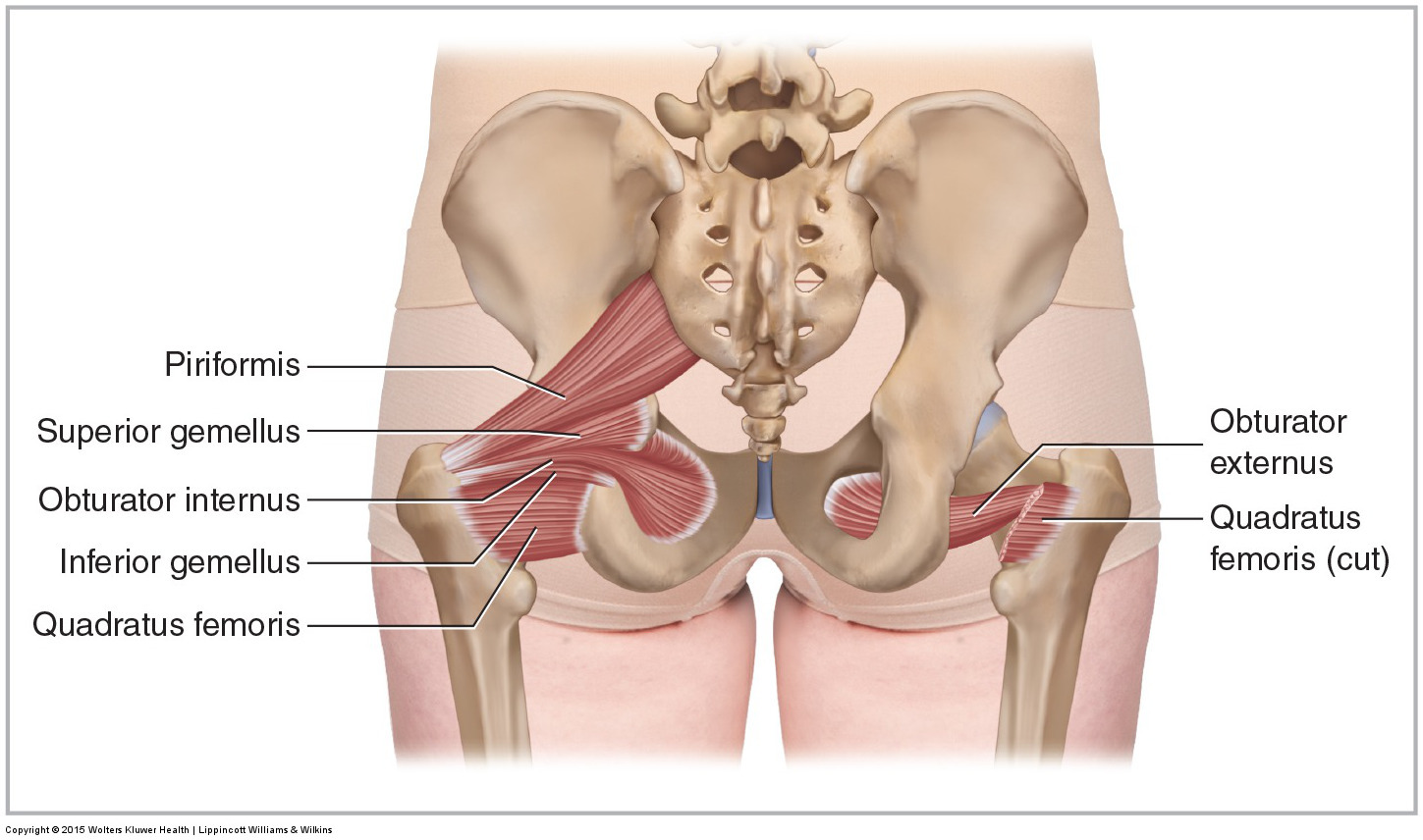
Muscles Of The Pelvis

Pelvis Anatomy Anatomy Bones Hip Anatomy
Pelvis And Perineum Basicmedical Key

Three Dimensional Posterior View Of The Pelvis Download Scientific Diagram
0 comments
Post a Comment